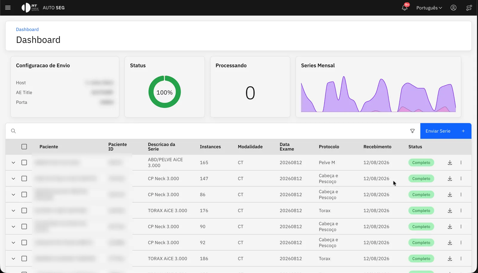
Automatic contours, straight into your TPS.

Models trained on real clinical data return a standard RTSTRUCT to your planning system — no scripts, no operator.

44AI models in the catalog
348TG-263 structures
3interface languages
0clicks from scanner to TPS
How it works

From the DICOM received to the RTSTRUCT delivered — one platform

Tabs advance automatically; hover or focus the card to pause.

A contouring workstation in the browser

Open the case in any browser — macOS, Windows, Linux or a tablet — and refine the AI contours with the tools a planner expects, on the CT itself.

  • Brush, 3D sphere brush, threshold brush, flood fill and eraser — 1 to 50 mm radius
  • Slice interpolation between the cuts you draw
  • 3D margin, HU threshold and structure algebra (union, subtract, intersect)
  • Axial, sagittal, coronal and multi-axial layouts, plus a 3D surface view
  • Contour versions with physician approval
1–50 mmbrush radius, 2D and 3D sphere

Editor brush settings

This is an interactive demo. Arrow keys move between tools, Tab reaches each structure, and nothing you do here leaves your browser.

AutoSegLCTSC-Test-S3-104

Studies

LCTSC-Test-S3-104
CT study · 1.3.6.1.4.1…
Thorax
RTSTRUCT 1

ROI Workspace

Organs at risk
Organ
Organ
Organ
Organ
Organ

Selected ROI

ID3
NameHeart
TypeOrgan
StatusUnapproved
Color#d63031
Volume677 cm³
Heart
LCTSC-Test-S3-104
CT · Im: 92/165
Heart
AXIALWW: 400 WL: 40
Zoom: 1.00x
APRL

Tool settings · Crosshairs

Drag the crosshair center in any viewport to synchronize the other planes.

Interactions

Left drag
Crosshairs
Shift + left drag
Window/Level
Right drag
Pan
Middle drag
Zoom
Wheel
Stack scroll

Versions

Commit message

Commit

No versions yet — save or commit to create one.

Structures

348 TG-263 structures across eight regions

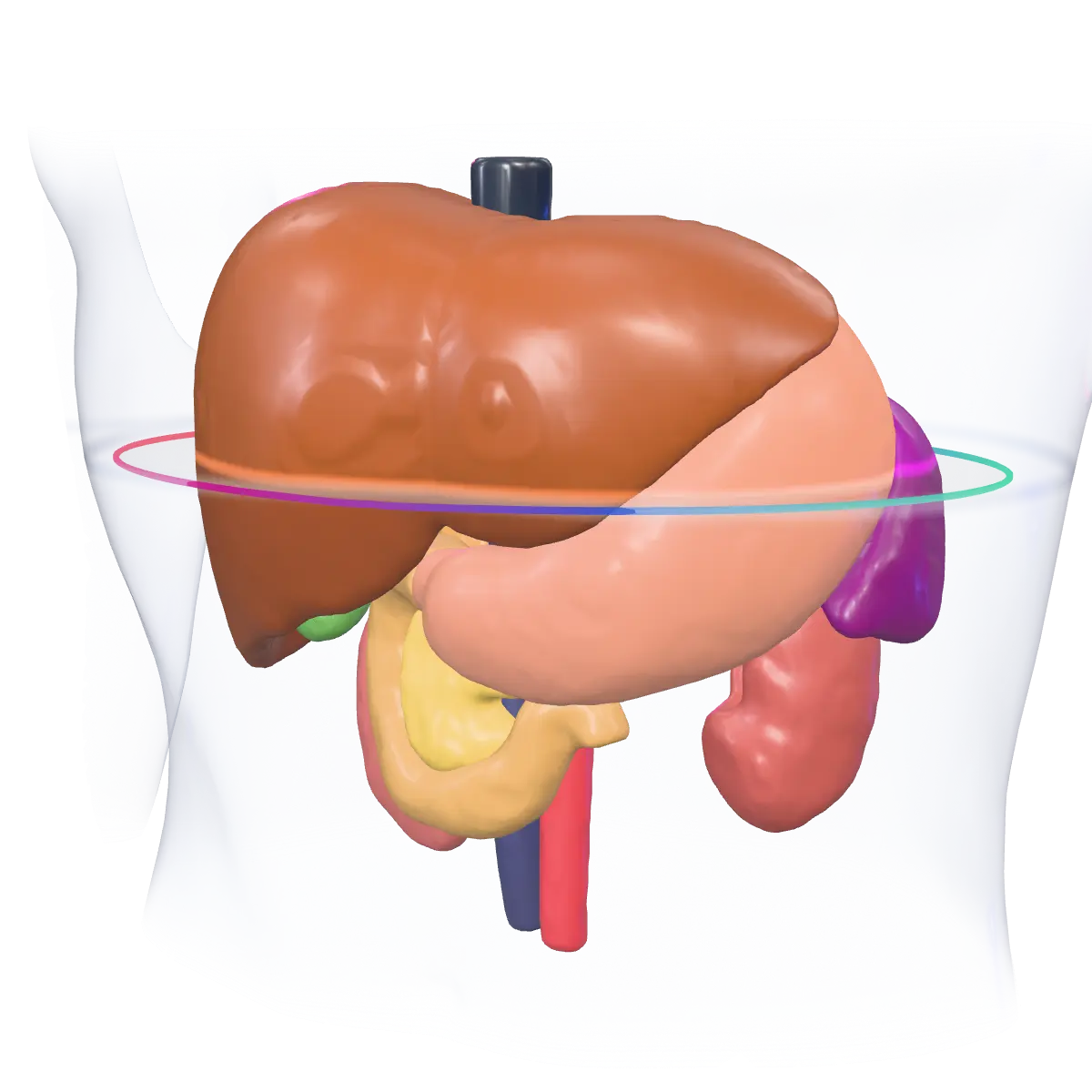
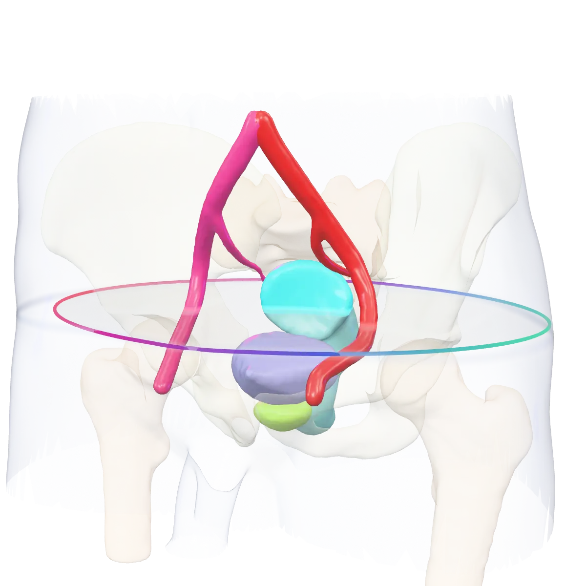
Each region below shows what AutoSeg segments today — the same names and colours you will see in the viewer and in the RTSTRUCT. Browse by region, or search the full list.

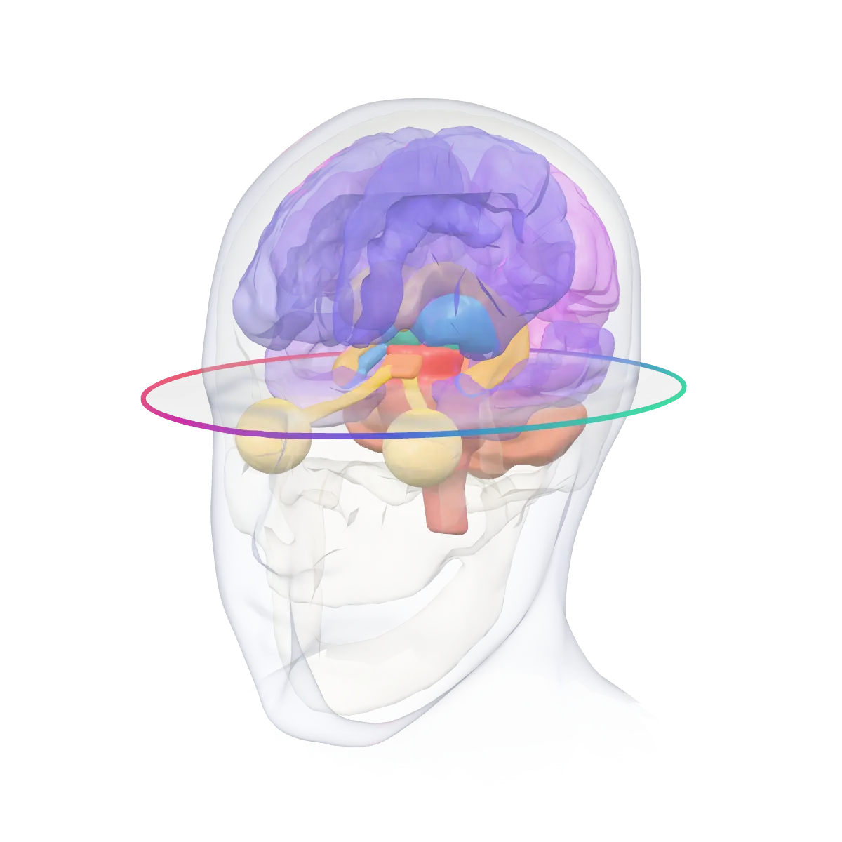
  • Head & Neck

    From the brainstem to the larynx

    Parotid and submandibular glands, brainstem and spinal cord, optic nerves, chiasm and lenses, cochleae, oral cavity, pharyngeal constrictors, mandible, thyroid and larynx — the organs at risk of a head and neck plan, each side named apart.

    125TG-263 structures
    Three-dimensional head and neck from a public atlas: brain, eyes, optic nerves, submandibular glands, mandible, tongue and larynx inside a translucent head, coloured the way AutoSeg colours them.
    Anatomical model · public atlases
    • A_Carotid_L
    • A_Carotid_R
    • A_IntCarotid_L
    • A_IntCarotid_R
    • AntScalene_L
    • AntScalene_R
    • Arytenoid
    • AudCanal_L
    • AudCanal_R
    • Bone_Mandible
    • BrachialPlex_L
    • BrachialPlex_R
    • BrachialPlexi
    • Brain
    • BuccalMucosa
    • Chiasm
    • Cochlea_L
    • Cochlea_R
    • Cochleae
    • CricoidCart
    • Cricopharyngeus
    • Digastric_L
    • Digastric_R
    • Esophagus_S
    • Eye_AL
    • Eye_AR
    • Eye_L
    • Eye_PL
    • Eye_PR
    • Eye_R
    • Face
    • Glnd_Lacrimal_L
    • Glnd_Lacrimal_R
    • Glottis
    • HardPalate
    • Head
    • Hyoid
    • Hypopharynx
    • InfOblique_L
    • InfOblique_R
    • InfRect_L
    • InfRect_R
    • Larynx
    • Larynx_SG
    • LarynxAir
    • LatPterygoid_L
    • LatPterygoid_R
    • LatRect_L
    • LatRect_R
    • Lens_L
    • Lens_R
    • LevPalpebrae_L
    • LevPalpebrae_R
    • LevScap_L
    • LevScap_R
    • Lips
    • Mandible
    • Masseter_L
    • Masseter_R
    • MedPterygoid_L
    • MedPterygoid_R
    • MedRect_L
    • MedRect_R
    • MidScalene_L
    • MidScalene_R
    • Mucosa
    • NasalCav_L
    • NasalCav_R
    • Nasopharynx
    • OpticNerve_L
    • OpticNerve_R
    • OpticNerves
    • OralCavity
    • Oropharynx
    • Parotid_L
    • Parotid_R
    • Parotids
    • PharConstr_I
    • PharConstr_M
    • PharConstr_S
    • Pharynx
    • Pituitary
    • Platysma_L
    • Platysma_R
    • PostScalene_L
    • PostScalene_R
    • Prevertebral_L
    • Prevertebral_R
    • SCM_L
    • SCM_R
    • Sinus_Frontal
    • Sinus_Max
    • Skull
    • SoftPalate
    • SpinalCanal
    • Sternothyroid_L
    • Sternothyroid_R
    • Styloid_L
    • Styloid_R
    • Submand_L
    • Submand_R
    • Submandibular_L
    • Submandibular_R
    • Submandibulars
    • SupOblique_L
    • SupOblique_R
    • SupRect_L
    • SupRect_R
    • Teeth_Lower
    • Teeth_Upper
    • Temporalis_L
    • Temporalis_R
    • TemporalLobe_L
    • TemporalLobe_R
    • Thyrohyoid_L
    • Thyrohyoid_R
    • Thyroid
    • ThyroidCart
    • Tongue
    • Trapezius_L
    • Trapezius_R
    • V_IntJugular_L
    • V_IntJugular_R
    • Zygomatic_L
    • Zygomatic_R

Full list

348 of 348 structures

Brain43

  • Amygdala_LAmygdala (L)
  • Amygdala_RAmygdala (R)
  • BloodBlood Vessels (Head)
  • Bone_CompactCompact Bone
  • Bone_SpongySpongy Bone
  • Brain_GMGray Matter
  • Brain_WMWhite Matter
  • BrainstemBrainstem
  • Caudate_LCaudate Nucleus (L)
  • Caudate_RCaudate Nucleus (R)
  • CerebellumCerebellum
  • Cingulate_LCingulate Cortex (L)
  • Cingulate_RCingulate Cortex (R)
  • CSFCerebrospinal Fluid
  • EyesEyeballs (Both)
  • FourthVentricleFourth Ventricle
  • Frontal_LFrontal Lobe (L)
  • Frontal_RFrontal Lobe (R)
  • Hippocampus_LHippocampus (L)
  • Hippocampus_RHippocampus (R)
  • Insula_LInsula (L)
  • Insula_RInsula (R)
  • LateralVentricle_LLateral Ventricle (L)
  • LateralVentricle_RLateral Ventricle (R)
  • Musc_HeadHead Muscles
  • NucleusAccumbens_LNucleus Accumbens (L)
  • NucleusAccumbens_RNucleus Accumbens (R)
  • Occipital_LOccipital Lobe (L)
  • Occipital_ROccipital Lobe (R)
  • Pallidum_LGlobus Pallidus (L)
  • Pallidum_RGlobus Pallidus (R)
  • Parietal_LParietal Lobe (L)
  • Parietal_RParietal Lobe (R)
  • Putamen_LPutamen (L)
  • Putamen_RPutamen (R)
  • ScalpScalp
  • Temporal_LTemporal Lobe (L)
  • Temporal_RTemporal Lobe (R)
  • Thalamus_LThalamus (L)
  • Thalamus_RThalamus (R)
  • ThirdVentricleThird Ventricle
  • VentralDC_LVentral Diencephalon (L)
  • VentralDC_RVentral Diencephalon (R)

Head & Neck125

  • A_Carotid_LLeft Common Carotid Artery
  • A_Carotid_RRight Common Carotid Artery
  • A_IntCarotid_LLeft Internal Carotid Artery
  • A_IntCarotid_RRight Internal Carotid Artery
  • AntScalene_LLeft Anterior Scalene
  • AntScalene_RRight Anterior Scalene
  • ArytenoidArytenoid
  • AudCanal_LLeft Auditory Canal
  • AudCanal_RRight Auditory Canal
  • Bone_MandibleMandible
  • BrachialPlex_LBrachial Plexus (L)
  • BrachialPlex_RBrachial Plexus (R)
  • BrachialPlexiBrachial Plexi (Both)
  • BrainBrain
  • BuccalMucosaBuccal Mucosa
  • ChiasmOptic Chiasm
  • Cochlea_LCochlea (L)
  • Cochlea_RCochlea (R)
  • CochleaeCochleae (Both)
  • CricoidCartCricoid Cartilage
  • CricopharyngeusCricopharyngeus
  • Digastric_LLeft Digastric
  • Digastric_RRight Digastric
  • Esophagus_SCervical Esophagus
  • Eye_ALEye Anterior Segment (L)
  • Eye_AREye Anterior Segment (R)
  • Eye_LLeft Eye
  • Eye_PLEye Posterior Segment (L)
  • Eye_PREye Posterior Segment (R)
  • Eye_RRight Eye
  • FaceFace
  • Glnd_Lacrimal_LLacrimal Gland (L)
  • Glnd_Lacrimal_RLacrimal Gland (R)
  • GlottisGlottis
  • HardPalateHard Palate
  • HeadHead
  • HyoidHyoid Bone
  • HypopharynxHypopharynx
  • InfOblique_LLeft Inferior Oblique Muscle
  • InfOblique_RRight Inferior Oblique Muscle
  • InfRect_LLeft Inferior Rectus Muscle
  • InfRect_RRight Inferior Rectus Muscle
  • LarynxLarynx
  • Larynx_SGSupraglottic Larynx
  • LarynxAirLaryngeal Airway
  • LatPterygoid_LLeft Lateral Pterygoid
  • LatPterygoid_RRight Lateral Pterygoid
  • LatRect_LLeft Lateral Rectus Muscle
  • LatRect_RRight Lateral Rectus Muscle
  • Lens_LLeft Eye Lens
  • Lens_RRight Eye Lens
  • LevPalpebrae_LLeft Levator Palpebrae
  • LevPalpebrae_RRight Levator Palpebrae
  • LevScap_LLeft Levator Scapulae
  • LevScap_RRight Levator Scapulae
  • LipsLips
  • MandibleMandible
  • Masseter_LLeft Masseter
  • Masseter_RRight Masseter
  • MedPterygoid_LLeft Medial Pterygoid
  • MedPterygoid_RRight Medial Pterygoid
  • MedRect_LLeft Medial Rectus Muscle
  • MedRect_RRight Medial Rectus Muscle
  • MidScalene_LLeft Middle Scalene
  • MidScalene_RRight Middle Scalene
  • MucosaOral Mucosa
  • NasalCav_LLeft Nasal Cavity
  • NasalCav_RRight Nasal Cavity
  • NasopharynxNasopharynx
  • OpticNerve_LLeft Optic Nerve
  • OpticNerve_RRight Optic Nerve
  • OpticNervesOptic Nerves (Both)
  • OralCavityOral Cavity
  • OropharynxOropharynx
  • Parotid_LLeft Parotid Gland
  • Parotid_RRight Parotid Gland
  • ParotidsParotid Glands (Both)
  • PharConstr_IInferior Pharyngeal Constrictor
  • PharConstr_MMiddle Pharyngeal Constrictor
  • PharConstr_SSuperior Pharyngeal Constrictor
  • PharynxPharynx
  • PituitaryPituitary Gland
  • Platysma_LLeft Platysma
  • Platysma_RRight Platysma
  • PostScalene_LLeft Posterior Scalene
  • PostScalene_RRight Posterior Scalene
  • Prevertebral_LLeft Prevertebral
  • Prevertebral_RRight Prevertebral
  • SCM_LLeft Sternocleidomastoid
  • SCM_RRight Sternocleidomastoid
  • Sinus_FrontalFrontal Sinus
  • Sinus_MaxMaxillary Sinus
  • SkullSkull
  • SoftPalateSoft Palate
  • SpinalCanalSpinal Canal
  • Sternothyroid_LLeft Sternothyroid
  • Sternothyroid_RRight Sternothyroid
  • Styloid_LLeft Styloid Process
  • Styloid_RRight Styloid Process
  • Submand_LLeft Submandibular Gland
  • Submand_RRight Submandibular Gland
  • Submandibular_LSubmandibular Gland (L)
  • Submandibular_RSubmandibular Gland (R)
  • SubmandibularsSubmandibular Glands (Both)
  • SupOblique_LLeft Superior Oblique Muscle
  • SupOblique_RRight Superior Oblique Muscle
  • SupRect_LLeft Superior Rectus Muscle
  • SupRect_RRight Superior Rectus Muscle
  • Teeth_LowerLower Teeth
  • Teeth_UpperUpper Teeth
  • Temporalis_LLeft Temporalis
  • Temporalis_RRight Temporalis
  • TemporalLobe_LTemporal Lobe (L)
  • TemporalLobe_RTemporal Lobe (R)
  • Thyrohyoid_LLeft Thyrohyoid
  • Thyrohyoid_RRight Thyrohyoid
  • ThyroidThyroid Gland
  • ThyroidCartThyroid Cartilage
  • TongueTongue
  • Trapezius_LLeft Trapezius
  • Trapezius_RRight Trapezius
  • V_IntJugular_LLeft Internal Jugular Vein
  • V_IntJugular_RRight Internal Jugular Vein
  • Zygomatic_LLeft Zygomatic Arch
  • Zygomatic_RRight Zygomatic Arch

Thorax75

  • A_Aorta_AscAscending Aorta
  • A_Aorta_DscDescending Aorta
  • A_BrachiocephalicBrachiocephalic Trunk
  • A_PulmonaryPulmonary Artery
  • A_Subclavian_LLeft Subclavian Artery
  • A_Subclavian_RRight Subclavian Artery
  • AortaAorta
  • AtrialAppendage_LLeft Atrial Appendage
  • Atrium_LAtrium (L)
  • Atrium_RAtrium (R)
  • Bone_SternumSternum
  • Breast_LBreast (L)
  • Breast_RBreast (R)
  • Cavity_ThoraxThoracic Cavity
  • Chestwall_OARChest Wall
  • Clavicle_LLeft Clavicle
  • Clavicle_RRight Clavicle
  • CostalCartilagesCostal Cartilages
  • EsophagusEsophagus
  • HeartHeart
  • Implant_BreastBreast Implant
  • LatDorsi_LLeft Latissimus Dorsi
  • LatDorsi_RRight Latissimus Dorsi
  • Lung_LLeft Lung
  • Lung_L_LLLeft Lung Lower Lobe
  • Lung_L_ULLeft Lung Upper Lobe
  • Lung_RRight Lung
  • Lung_R_LLRight Lung Lower Lobe
  • Lung_R_MLRight Lung Middle Lobe
  • Lung_R_ULRight Lung Upper Lobe
  • LungAirwaysLung Airways
  • LungAirwaysWallLung Airways Wall
  • LungArteriesLung Arteries
  • LungVeinsLung Veins
  • MediastinumMediastinum
  • MyocardiumMyocardium
  • PecMajor_LLeft Pectoralis Major
  • PecMajor_RRight Pectoralis Major
  • PericardiumPericardium
  • Rib_L_1Left Rib 1
  • Rib_L_2Left Rib 2
  • Rib_L_3Left Rib 3
  • Rib_L_4Left Rib 4
  • Rib_L_5Left Rib 5
  • Rib_L_6Left Rib 6
  • Rib_L_7Left Rib 7
  • Rib_L_8Left Rib 8
  • Rib_L_9Left Rib 9
  • Rib_L_10Left Rib 10
  • Rib_L_11Left Rib 11
  • Rib_L_12Left Rib 12
  • Rib_R_1Right Rib 1
  • Rib_R_2Right Rib 2
  • Rib_R_3Right Rib 3
  • Rib_R_4Right Rib 4
  • Rib_R_5Right Rib 5
  • Rib_R_6Right Rib 6
  • Rib_R_7Right Rib 7
  • Rib_R_8Right Rib 8
  • Rib_R_9Right Rib 9
  • Rib_R_10Right Rib 10
  • Rib_R_11Right Rib 11
  • Rib_R_12Right Rib 12
  • Scapula_LLeft Scapula
  • Scapula_RRight Scapula
  • SerratusAnt_LLeft Serratus Anterior
  • SerratusAnt_RRight Serratus Anterior
  • SternumSternum
  • TracheaTrachea
  • V_Brachiocephalic_LLeft Brachiocephalic Vein
  • V_Brachiocephalic_RRight Brachiocephalic Vein
  • V_Cava_SSuperior Vena Cava
  • V_PulmonaryPulmonary Vein
  • Ventricle_LVentricle (L)
  • Ventricle_RVentricle (R)

Abdomen23

  • Adrenal_LLeft Adrenal Gland
  • Adrenal_RRight Adrenal Gland
  • Cavity_AbdAbdominal Cavity
  • ColonColon
  • DuodenumDuodenum
  • ExtOblique_LLeft External Oblique
  • ExtOblique_RRight External Oblique
  • GallbladderGallbladder
  • IntOblique_LLeft Internal Oblique
  • IntOblique_RRight Internal Oblique
  • Kidney_LLeft Kidney
  • Kidney_RRight Kidney
  • KidneyCyst_LLeft Kidney Cyst
  • KidneyCyst_RRight Kidney Cyst
  • LiverLiver
  • PancreasPancreas
  • PortalVeinPortal Vein
  • RectusAbd_LLeft Rectus Abdominis
  • RectusAbd_RRight Rectus Abdominis
  • SmallBowelSmall Bowel
  • SpleenSpleen
  • StomachStomach
  • V_Cava_IInferior Vena Cava

Pelvis38

  • A_Iliac_LLeft Iliac Artery
  • A_Iliac_RRight Iliac Artery
  • BladderUrinary Bladder
  • Bowel_BagBowel Bag
  • CaudaEquinaCauda Equina
  • Colon_SigmoidSigmoid Colon
  • Femur_LLeft Femur
  • Femur_RRight Femur
  • Glut_Max_LLeft Gluteus Maximus
  • Glut_Max_RRight Gluteus Maximus
  • Glut_Med_LLeft Gluteus Medius
  • Glut_Med_RRight Gluteus Medius
  • Glut_Min_LLeft Gluteus Minimus
  • Glut_Min_RRight Gluteus Minimus
  • IliacA_LLeft Iliac Artery
  • IliacA_RRight Iliac Artery
  • IliacV_LLeft Iliac Vein
  • IliacV_RRight Iliac Vein
  • Iliopsoas_LLeft Iliopsoas
  • Iliopsoas_RRight Iliopsoas
  • PelvicBone_LLeft Pelvic Bone
  • PelvicBone_RRight Pelvic Bone
  • Pelvis_LLeft Hip
  • Pelvis_RRight Hip
  • PenileBulbPenile Bulb
  • ProstateProstate
  • Psoas_LLeft Psoas Major
  • Psoas_RRight Psoas Major
  • QuadLumb_LLeft Quadratus Lumborum
  • QuadLumb_RRight Quadratus Lumborum
  • RectumRectum
  • SacralPlex_LSacral Plexus (L)
  • SacralPlex_RSacral Plexus (R)
  • SacrumSacrum
  • SeminalVesSeminal Vesicles
  • UteroCervixUterus & Cervix
  • V_Iliac_LLeft Iliac Vein
  • V_Iliac_RRight Iliac Vein

Spine35

  • Autochthon_LLeft Autochthonous Muscle
  • Autochthon_RRight Autochthonous Muscle
  • C1Vertebra C1
  • C2Vertebra C2
  • C3Vertebra C3
  • C4Vertebra C4
  • C5Vertebra C5
  • C6Vertebra C6
  • C7Vertebra C7
  • ErectorSpinae_LLeft Erector Spinae
  • ErectorSpinae_RRight Erector Spinae
  • IVDIntervertebral Discs
  • L1Vertebra L1
  • L2Vertebra L2
  • L3Vertebra L3
  • L4Vertebra L4
  • L5Vertebra L5
  • Paraspinal_LLeft Paraspinal Muscle
  • Paraspinal_RRight Paraspinal Muscle
  • S1Vertebra S1
  • SpinalCordSpinal Cord
  • T1Vertebra T1
  • T2Vertebra T2
  • T3Vertebra T3
  • T4Vertebra T4
  • T5Vertebra T5
  • T6Vertebra T6
  • T7Vertebra T7
  • T8Vertebra T8
  • T9Vertebra T9
  • T10Vertebra T10
  • T11Vertebra T11
  • T12Vertebra T12
  • Transversospinalis_LLeft Transversospinalis
  • Transversospinalis_RRight Transversospinalis

Extremities2

  • Humerus_LLeft Humerus
  • Humerus_RRight Humerus

Whole body7

  • BodyBody
  • BodyExtremBody Extremities
  • BodyTruncBody Trunk
  • BonesBones (All)
  • GlandsGlands (All)
  • MusclesMuscle (All)
  • Tissue_SubcutSubcutaneous Tissue
Our own models

We train our own models

AutoSeg's specialist models are trained and maintained by the RT Medical Systems team on our own GPU infrastructure — male and female pelvis, neural structures, brain MR and more. OAR coverage with TG-263 nomenclature across head & neck, thorax, abdomen, pelvis and spine.

RT Medical Systems logoNVIDIA Inception program badge

RT Medical Systems is a member of the NVIDIA Inception program.

30nnU-Net models trained in-house
348distinct structures in the catalog
CT + MRinput modalities
autoseg.rtmedical.ai
Brain MR slices with contoured cerebral structures and the reconstructed 3D brain mesh in AutoSeg
Expert reference case

Scroll a chest CT the way a planner does

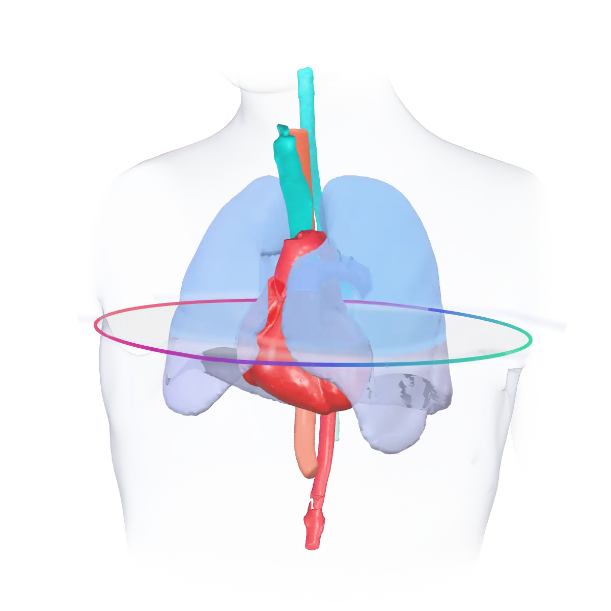
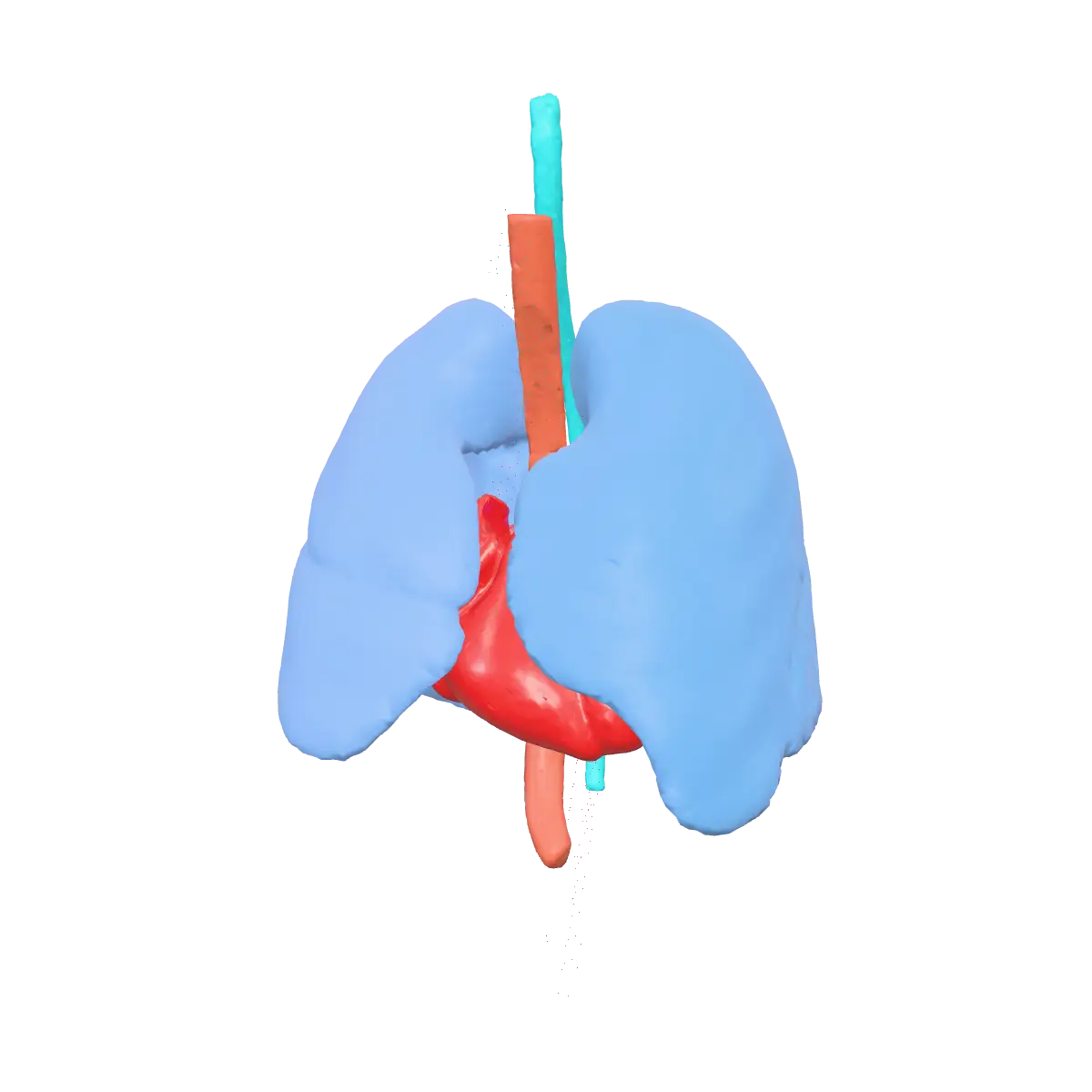
1 cuts through one planning CT, from the lung apices to the bases: both lungs, heart, esophagus and spinal cord. Every mask here was drawn voxel by voxel by experts for a public reference dataset — the ground truth an auto-segmentation is judged against.

—slice
Runs where you are

Multi-hospital, on your terms

Isolation per hospital, federated identities and the same product wherever it is installed — with two people on the same case when the clinic needs it.

Multi-hospital with SSO

Per-hospital isolation, role-based access control, and corporate federation via SAML, OIDC and LDAP — with an immutable audit trail of every event.

Cloud, on-premise or air-gapped

The same product, packaged for Kubernetes/Helm: cloud SaaS, a fully in-hospital installation, or an isolated environment with no internet at all.

Four-handed contouring

2 people on this study

Per-structure locks and live presence: the dosimetrist draws the OARs while the physician draws the PTV — same patient, same time. Each sees where the other is working, and every edit is recorded with author and sequence.

TPS integration

Closes the loop with Eclipse and Monaco — over pure DICOM

No ESAPI, no scripting, no plugins. AutoSeg speaks C-STORE/C-ECHO and delivers standard RTSTRUCT with TG-263 nomenclature — compatible with Varian Eclipse, Elekta Monaco, RaySearch RayStation, and any TPS that speaks DICOM.

Return delivery is handled by the AutoSeg Desktop agent, a lightweight service running inside the hospital network — clinical data never has to leave the institution.

  • Varian Eclipse
  • Elekta Monaco
  • RaySearch RayStation
  • any DICOM TPS
  1. 01

    The scan is done

    The images travel from the scanner to AutoSeg on their own — nothing to export, nobody to ask.

  2. 02

    Contours drawn automatically

    The organs are outlined in minutes, inside the hospital's network or in the cloud.

  3. 03

    Ready in your planning system

    The structures appear in your planning system with the names and colours your team already uses.

  4. 04

    Reviewed and approved by the physician

    The physician reviews, adjusts where needed and approves. Every version is kept.

The physician's approval goes back into the case history — nothing is ever overwritten.

Pricing

One variable only: segmentations per month

Transparent pricing with no licensing traps: you pay for the volume you process — and nothing else.

The only variable

segmentations / month

Pick the monthly volume tier that makes sense for your operation. It's the single number that defines your plan.

No counting linacs

We don't charge per linear accelerator, per user, per workstation, or per connected TPS. None of those numbers ever enter the bill.

  • per linac
  • per user
  • per workstation
  • per TPS

One queue, the same for everyone

No low-priority queue: a plan with fewer segmentations processes at the same speed and the same priority as the biggest volumes.

equal priority on every plan

Need a small volume? Perfect — start small and adjust the tier as your operation grows.

Size my volume
Compliance

Engineered for the regulated clinical environment

Software developed under medical-device (SaMD) processes, with risk management, traceability, and an immutable audit trail.

  • ISO 13485
  • IEC 62304 Class C
  • ISO 14971
  • LGPD
  • HIPAA
  • ANVISA RDC 657/2022
  • TG-263

See AutoSeg running on your workflow

Schedule a demo with our team — or open the application right now.